Melanoma Coroidale
Melanoma is a kind of cancer that begins in the skin cells that create the pigment melaninMelanoma usually starts as a dark mole on the skin However, it can also form in other tissue, such as.

Melanoma coroidale. Plaque radiation or externalbeam radiation), transpupillary thermotherapy (TTT), local resection, or enucleation Large tumors warrant enucleation. The most common site for ocular melanoma is the choroid, followed by the ciliary body, and the iris In the United States, choroidal melanoma affects approximately 2500 people per year with an incidence of 6 per million Ocular melanoma is more common in fairskinned individuals. The prognosis of choroidal melanoma is based on clinical, histologic, and genetic parameters (see Chapter 141, Molecular genetics of choroidal melanoma) Some parameters such as ciliary body invasion, extraocular extension and the largest basal diameter are incorporated into the TNM classification 9 Histopathologic examination allows for.
Choroidal melanoma is the most common primary malignant intraocular tumor with an annual incidence in the USA of 08 cases per 100 000 population 1 Once metastasis becomes clinically apparent, the 1year mortality rate approaches 80% 2 Given this poor prognosis, enucleation was historically considered the only appropriate management for. Choroidal melanomas Treating melanomas that start in the choroid depends on the size of the tumor and how well the eye functions The smaller the tumor, the less likely surgery will be needed, unless the eye is badly damaged or vision is lost Small melanomas There are often several options for treating small choroidal melanomas Both you and. Eye melanoma is a type of eye cancer Learn about symptoms and treatments for this rare cancer It's also called ocular melanoma or uveal melanoma COVID19 What you need to know Vaccine updates, safe care and visitor guidelines, and trusted coronavirus information.
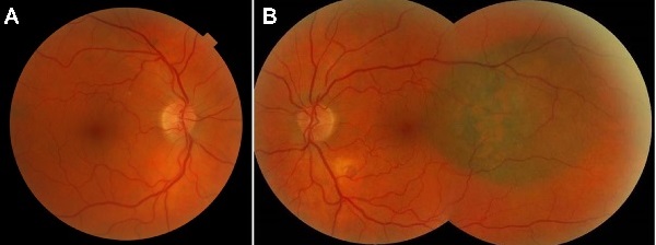
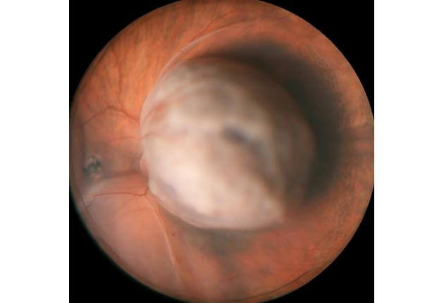
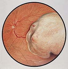
Choroidal melanoma is a disease with a high mortality rate, usually irrespective of the chosen treatment modality About 3050% of patients with choroidal melanoma will die within 10 years from. Choroidal melanoma is the most common primary malignant intraocular tumor with an annual incidence in the USA of 08 cases per 100 000 population 1 Once metastasis becomes clinically apparent, the 1year mortality rate approaches 80% 2 Given this poor prognosis, enucleation was historically considered the only appropriate management for. Choroidal melanoma is cancer found in the choroid This is a tissue that lines the eye between the retina and the sclera The sclera is the white part of your eye A choroidal melanoma can become large and cause your retina to detach This can cause vision loss.
Metastasis is common following primary diagnosis of choroidal melanoma Patterns of metastatic spread have been described Frequently the site of metastasis is the liver, with the proportion of cases with liver metastasis ranging from 56% to 100% 1 8 Diagnosis of metastasis is often followed by rapid physical decline of the patient. Choroidal melanoma treatments and varying rates of success as this is a rather aggressive and fast spreading cancer that often goes undetected for too long Immunotherapy drugs are being tested, but currently pembrolizumab (Keytruda) and ipilmumad (Yervoy) are heavily immplemented Brachytherapy (Plaque therapy) is when doctors plant seeds of. A choroidal nevus is a flat, benign pigmented area that appears in the back of the eye and is basically an eye freckle If your doctor refers to a lesion in your eye that needs to be tracked, she is most likely talking about a choroidal nevus The accumulation of pigmented cells that makes up a nevus occurs in a very small percentage of the population.
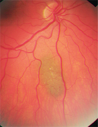
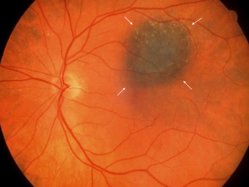
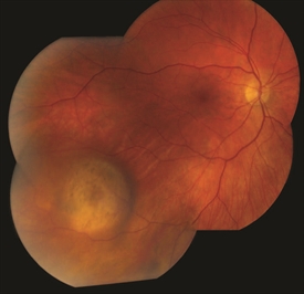
Melanoma is a type of cancer that develops in the cells that produce melanin — the pigment that gives your skin its color Your eyes also have melaninproducing cells and can develop melanoma Eye melanoma is also called ocular melanoma Most eye melanomas form in the part of the eye you can't see when looking in a mirror. If the choroidal nevus has orange pigment or has thickened, it should be checked more often If a choroidal nevus is leaking subretinal fluid, this is a particularly ominous sign Such tumors should be followed most closely for evidence of growth or malignant transformation into a choroidal melanoma. Primary choroidal melanoma arises from melanocytes within the choroid It is thought to develop from preexisting melanocytic naevi, although de novo growth may occur The colour varies from darkly pigmented to amelanotic.
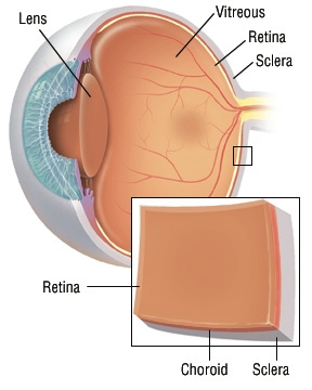
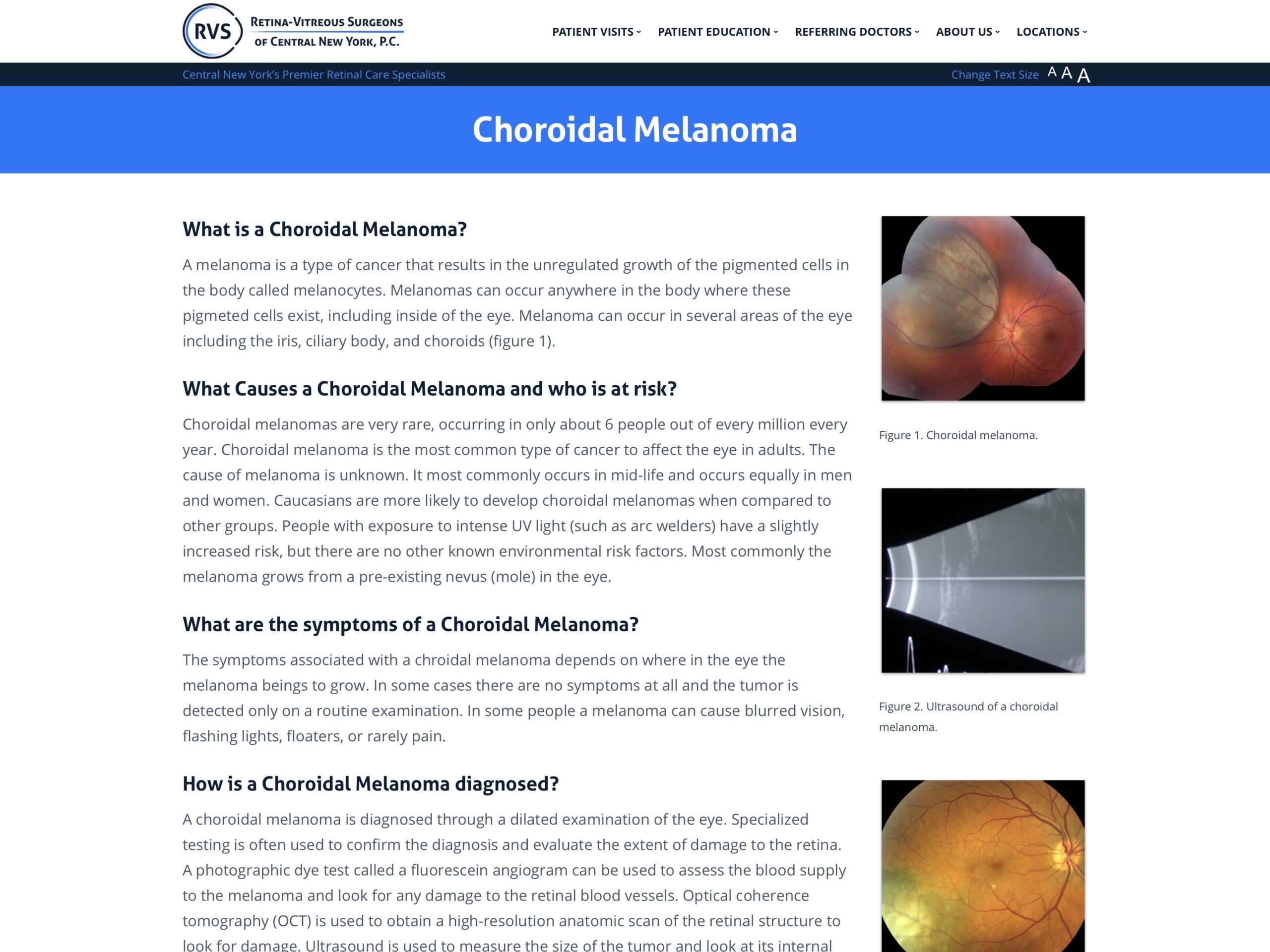
Choroidal melanoma is a cancer that affects part of the eye It develops in the choroid, the spongelike membrane at the back of the eye between the sclera (the white of the eye) and the retina (The retina is the lightsensitive structure at the back of the eye It sends visual information to the brain). Melanoma can occur in several regions of the eye including the iris, ciliary body, and choroid These regions are collectively known as the uvea It typically arises in midlife in both men and women with no environmental cause, except that it might be more common in arc welders. Choroidal melanoma is the most common type of intraocular melanoma The 5year relative survival rate for people with small choroidal melanoma is 84% The 5year relative survival rate for people with medium choroidal melanoma is 68% The 5year relative survival rate for people with large choroidal melanoma is 47% Ciliary body melanoma is.
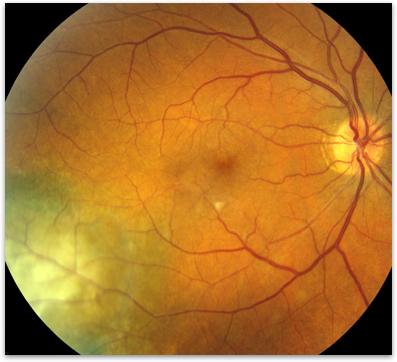
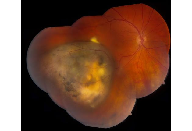
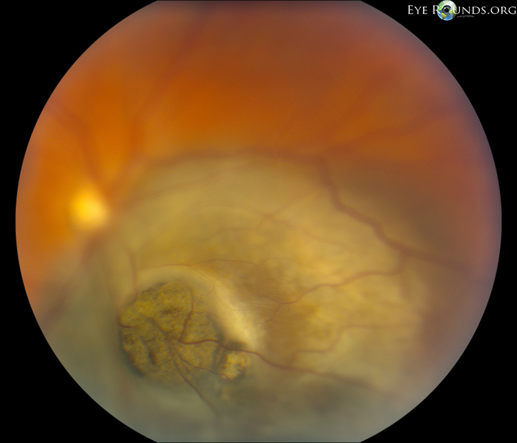
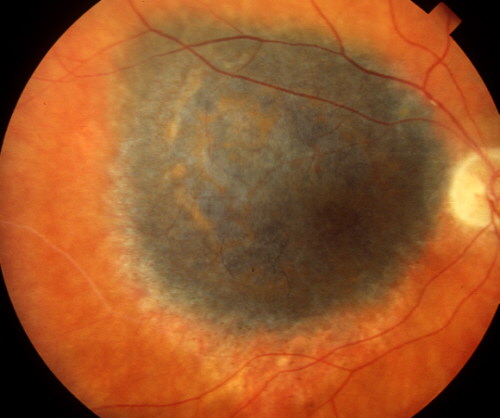
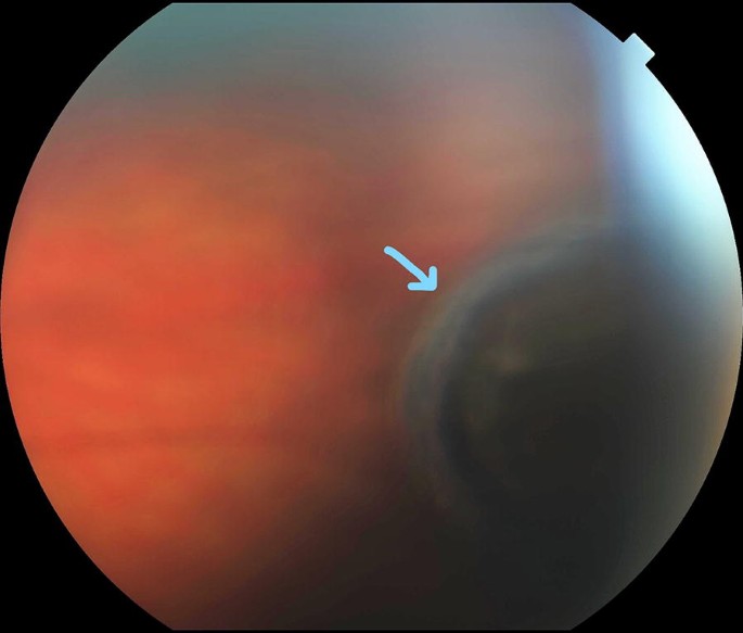
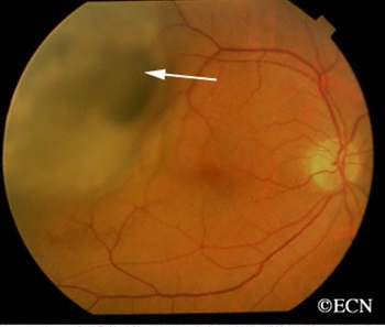
Choroidal Melanoma is a Life Sentence A thorough knowledge of treatment options and associated risks is crucial to ensure the best possible outcome with this dire condition By Sara Weidmayer, OD 1 A fundus photograph of our patient’s right eye. Choroidal melanomas Treating melanomas that start in the choroid depends on the size of the tumor and how well the eye functions The smaller the tumor, the less likely surgery will be needed, unless the eye is badly damaged or vision is lost Small melanomas There are often several options for treating small choroidal melanomas Both you and. N=55) were 77 mm (range –245) and 150 mm (range 29–250), respectively The corresponding numbers for group N were 50 mm (range –230) and 1 mm (range.
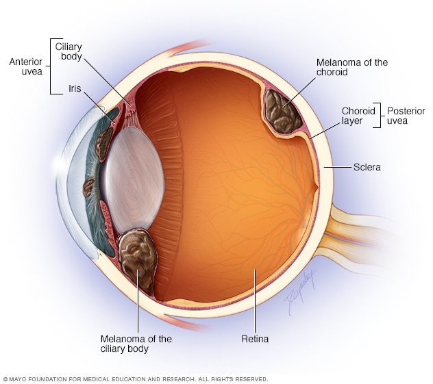
Choroidal melanoma is a subtype of uveal melanoma Uveal melanomas can be divided into 2 categories (1) anterior uveal melanomas, in which the tumor arises in the iris, and (2) posterior uveal melanomas, in which the tumor arises in either the choroid or the ciliary body. 1Choroidal nevi 2 Choroidal metastases 3 Choroidal hemangioma (hamartoma) Benign vascular lesion Circumscribed form in adults, sometimes with retinal detachment Diffuse form in infants associated with Sturge Weber Increased T2 signal and enhancement than melanoma 4. Choroidal Melanoma The wall of the eye has 3 main layers From outside to inside there is the white sclera, a blood vessel layer called the uvea (choroid, ciliary body and iris) and an inner retinal layer Further, the pigment producing cells, “melanocytes” are primarily found in the vascular uveal layer.
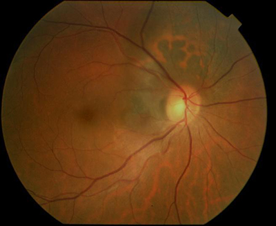
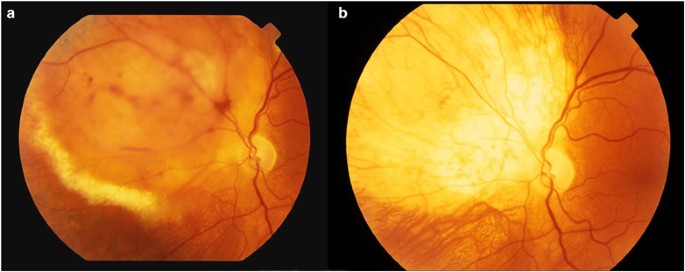
As an ocular melanoma arises from its benign counterpart called a choroidal nevus, it is important to differentiate a benign nevus from a highrisk nevus which may grow into a melanoma A variety of clinical features and parameters detected in a nevus may help determine whether a nevus or nevuslike lesion is at risk for growth or has already. A diagnosis of choroidal melanoma was made Treatment This was a mediumsized tumor (based on COMS) at the time of diagnosis Options for treatment included radiation (preferred therapy;. Choroidal nevi are benign melanocytic lesions of the posterior uvea In the United States, their prevalence ranges from 46 percent to 79 percent in Caucasians 1 By comparison, choroidal melanoma is rare, manifesting in approximately six in 1 million Caucasian individuals.
Plaque radiation or externalbeam radiation), transpupillary thermotherapy (TTT), local resection, or enucleation Large tumors warrant enucleation. A choroidal melanoma is a cancer that forms when malignant (cancerous) tumor cells grow inside the eye in the tissue layer called the choroid This layer of tissue lies beneath the retina and lines the inside of the back portion of the eye The choroid is the most common location for a melanoma inside the eye, but melanomas can also form inside. If the choroidal nevus has orange pigment or has thickened, it should be checked more often If a choroidal nevus is leaking subretinal fluid, this is a particularly ominous sign Such tumors should be followed most closely for evidence of growth or malignant transformation into a choroidal melanoma.
Choroidal Melanoma is the most common primary intraocular malignant tumor and the second most common intraocular tumor, behind metastases It is estimated that up to 50% of patients with choroidal melanoma develop metastatic disease, despite successful treatment of the primary tumor. What Is A Choroidal Nevus A choroidal nevus is a flat, benign pigmented area that appears in the back of the eye and is basically an eye freckle If your doctor refers to a lesion in your eye that needs to be tracked, she is most likely talking about a choroidal nevus. Malignant uveal melanomas, also referred to as choroidal melanomas, are the most common primary tumor of the adult eye 3.
Science and Education Publishing is an academic publisher of open access journals It also publishes academic books and conference proceedings SciEP currently has 100 open access journals in the areas of Earth &Environmental Sciences, Medicine &Healthcare, Biomedical &Life Sciences, Computer Science &Communications, Chemistry &Materials Science, Engineering, Physics &Mathematics, Business. Choroidal melanoma The median thickness and largest basal diameter of choroidal melanomas in group M (table 1;. Uveal melanoma is the most common primary intraocular malignancy in adults It arises from the neuroectodermal melanocytes within the uveal tract, most commonly involving the choroid (85%) (1) Choroidal melanoma presentation peaks at the age of 60 years with a higher incidence in Caucasians.
Choroidal melanoma is a cancer that affects part of the eye It develops in the choroid, the spongelike membrane at the back of the eye between the sclera (the white of the eye) and the retina (The retina is the lightsensitive structure at the back of the eye It sends visual information to the brain). Choroidal melanoma is cancer found in the choroid This is a tissue that lines the eye between the retina and the sclera The sclera is the white part of your eye A choroidal melanoma can become large and cause your retina to detach. Amelanotic melanoma is most recognizable by its reddish, pinkish, or nearly colorless look You may see a patch of abnormal skin but not the usual dark brown or black color that usually indicates.
The review also found small choroidal melanoma management typically involves plaque radiotherapy with five and 10year rates of tumor recurrence at 7% and 11%, visual acuity loss (≥3 Snellen lines) at 39% and 49% and melanomarelated metastasis at 4% and 9%, respectively. What is choroidal melanoma?. The most common site for ocular melanoma is the choroid, followed by the ciliary body, and the iris In the United States, choroidal melanoma affects approximately 2500 people per year with an incidence of 6 per million Ocular melanoma is more common in fairskinned individuals.
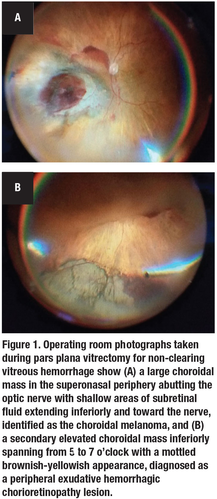
Choroidal melanoma is a cancer of the eye that develops in the pigmented cells of the choroid The choroid is composed of layers of blood vessels that nourish the back of the eye, located between the sclera and the retina It is unusual for a choroidal melanoma to spread to other parts of the body, but similar to skin melanoma, it is possible. Eye melanoma is a type of eye cancer Learn about symptoms and treatments for this rare cancer It's also called ocular melanoma or uveal melanoma COVID19 What you need to know Vaccine updates, safe care and visitor guidelines, and trusted coronavirus information. A Rare Presentation of Multifocal Choroidal Melanoma The multifocal lesions, themselves rare, were even more unexpected in the absence of ocular melanocytosis and germline BAP1 mutation.
Melanoma is a kind of cancer that begins in the skin cells that create the pigment melaninMelanoma usually starts as a dark mole on the skin However, it can also form in other tissue, such as. Choroidal melanoma – Choroid a coating of tissue that is present in the backside of the eye It helps to create a reflection, as it situated behind the retina and assists to produce a picture If uveal melanoma is initially started from the Choroid, then it termed as Choroidal melanomas and the most frequently found the ocular melanoma. A diagnosis of ocular melanoma can lead to grave outcomes, including enucleation and systemic spread Considering the potential progression of this condition, it is important for a practitioner to.
Purpose of review To review the current features and classification of choroidal melanoma, and to identify the lesions that clinically simulate choroidal melanoma (pseudomelanoma) Recent findings Uveal melanoma is a serious lifethreatening intraocular malignancy, most often found in Caucasians (98%) and primarily involving the choroid (90%), ciliary body (7%), or iris (2%). Choroidal Melanoma is a Life Sentence A thorough knowledge of treatment options and associated risks is crucial to ensure the best possible outcome with this dire condition By Sara Weidmayer, OD 1 A fundus photograph of our patient’s right eye. Unlike choroidal nevi, choroidal melanoma are exceedingly rarepresent at a rate of around 6 per 1 million individuals The overwhelming majority of choroidal nevi will never become a melanomao nly about one in over 8,000 nevi have a malignant transformation.
A 7yearold intact female golden Retriever was referred for evaluation of an intraorbital mass of the left eye Based on ophthalmoscopy, ultrasonography and magnetic resonance imaging (MRI), the tentative diagnosis was made as an intraocular neoplasia, especially choroidal melanoma The orbital exe. Choroidal melanoma is the second most common intraocular tumor – metastasis being the most frequent – and the most common primary intraocular malignancy in the adult population It arises from malignant uveal melanocytes. A choroidal melanoma is a cancer that forms when malignant (cancerous) tumor cells grow inside the eye in the tissue layer called the choroid This layer of tissue lies beneath the retina and lines the inside of the back portion of the eye The choroid is the most common location for a melanoma inside the eye, but melanomas can also form inside.
Uveal melanoma is a rare tumor with an incidence of 43 per million people in the general population 1 This malignancy is typically solitary and unilateral, rarely manifesting as multifocal unilateral or bilateral tumor 2 In an analysis of 8,033 eyes with uveal melanoma by Shields et al, less than 1% of patients (11 of 8,022) presented with either multifocal or bilateral tumors 2. Uveal melanoma is the leading primary eye cancer in adults Symptoms Melanoma can occur in several regions of the eye including the iris, ciliary body, and choroid These regions are collectively known as the uvea It typically arises in midlife in both men and women with no environmental cause, except that it might be more common in arc welders. Melanin is a pigment that gives your skin its color A change in your melanin color can often indicate that melanoma is developing in your skin With amelanotic melanoma, there isn’t always a.
Choroidal Melanoma Background Choroidal melanoma (CM) is the most common primary malignant intraocular tumour Patients are generally white, age 55–75, and have no known family history Local tumour control can be achieved in >90% of cases However, systemic metastasis occurs in 40% within 5 years of ocular treatment. The choroidal melanoma is malignant disorder, which arises from the melanin cells present in the eye The choroid is rich in vascular structure and therefore, the malignancy of the choroid melanoma usually not restricted to the eye, but it can spread to distant parts of the body Choroidal melanoma is the most common eye melanoma (1). A diagnosis of choroidal melanoma was made Treatment This was a mediumsized tumor (based on COMS) at the time of diagnosis Options for treatment included radiation (preferred therapy;.
Patient was operated subsequently, and histopathological diagnosis of choroidal melanoma was confirmed 2 article feature images from this case Primary uveal malignant melanoma. Choroidal melanoma treatments and varying rates of success as this is a rather aggressive and fast spreading cancer that often goes undetected for too long Immunotherapy drugs are being tested, but currently pembrolizumab (Keytruda) and ipilmumad (Yervoy) are heavily immplemented Brachytherapy (Plaque therapy) is when doctors plant seeds of. › Forums › Cutaneous Melanoma Community › Choroidal Nevus This topic has 1 reply, 1 voice, and was last updated 1 year, 10 months ago by María C Post.
Choroidal Melanoma Is A Life Sentence
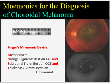
To Diagnose Choroidal Melanoma Just Remember Most New York Eye Cancer Center
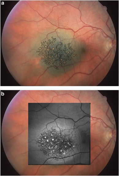
Review Of Fundus Autofluorescence In Choroidal Melanocytic Lesions Eye
Melanoma Coroidale のギャラリー

Choroidal Melanoma A Case Report Eyedocs

Choroidal Melanoma Symptoms Causes Treatment Options Retina Consultants Of Alabama

Small Choroidal Melanoma With Chromosome 3 Monosomy On Fine Needle Aspiration Biopsy Ophthalmology
Melanoma Or Pseudomelanoma

Choroidal Melanoma And Disclosing Bad News A Teaching Case Report The Journal Of Optometric Education

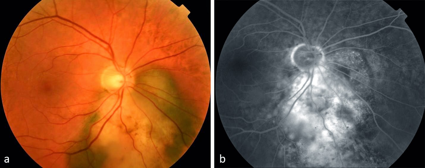
Preretinal Neovascularisation Associated With Choroidal Melanoma British Journal Of Ophthalmology

Plaque Placement For A Choroidal Melanoma The Medical University Of South Carolina
.jpg)
Choroidal Melanoma Cancer Choroidal Melanoma Eye Cancer Treatment In Mumbai India
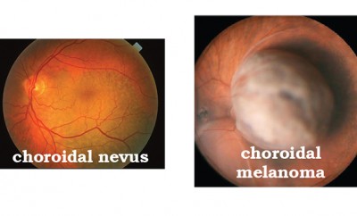
Ocular Tumors Choroidal Nevus Vs Choroidal Melanoma The Women S Journal
Optician
Choroidal Melanoma Harvard Health

Atypical Choroidal Melanoma Report Of 3 Cases
Choroidal Melanoma And Disclosing Bad News Br A Teaching Case Report Br
Q Tbn And9gcs Warivbt5u1fx1h9tibx Ca5wt5gplo2mnfz00eeb57zbbdyk Usqp Cau

Local Recurrence Of Choroidal Melanoma In The Right Eye Patient Had Download Scientific Diagram
Choroidal Melanoma Retina Vitreous Surgeons Of Cny
Pitfalls In The Diagnosis Of Choroidal Tumors 3 Case Reports Of Choroidal Masses
Choroidal Melanoma Europe American Academy Of Ophthalmology
Small Choroidal Melanoma To Treat Or Not To Treat That Is The Question New York Eye Cancer Center
Choroidal Melanoma Affecting The Optic Nerve New York Eye Cancer Center
Choroidal Melanoma Wills Eye Hospital
Managing Choroidal Melanomas
Q Tbn And9gcrrdklshyh2oqz5ffhgijjy1b9exifn500nzztrszdgrl3shpq4 Usqp Cau

Omf Basics Of Om

Retinal Physician The Treatment Of Choroidal Melanoma How Why When And By Whom

Bilateral Breast Metastases From Choroidal Melanoma American Journal Of Ophthalmology
Choroidal Melanoma
Eye Melanoma Symptoms And Causes Mayo Clinic

Choroidal Melanoma
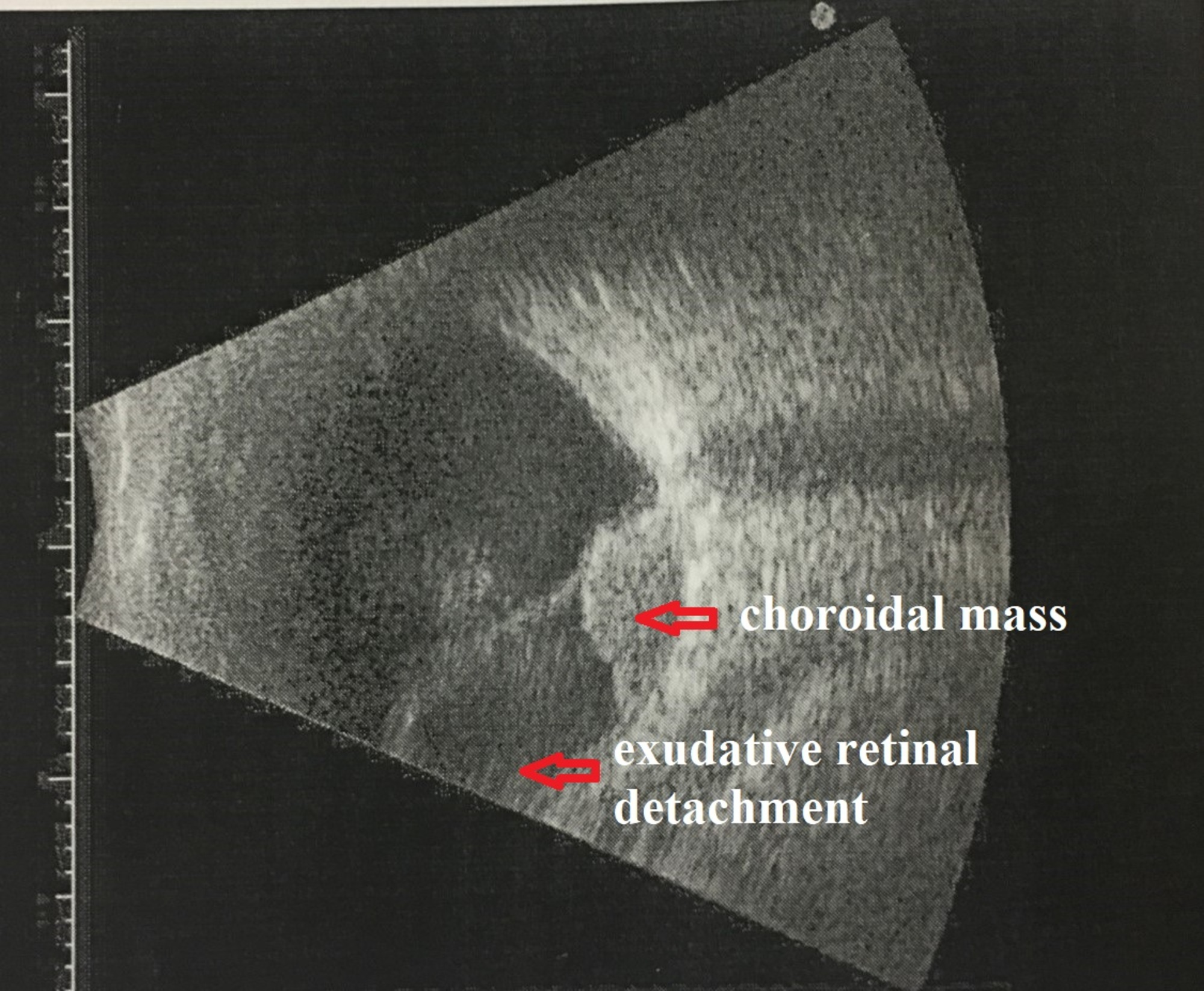
Choroidal Malignant Melanoma 58 Year Old Female With Pigmented Retinal Lesion And Exudative Retinal Detachment

Retinal Physician An Update On Intravitreal Chemotherapy

Large Choroidal Melanoma Retina Image Bank

Amelanotic Choroidal Melanoma Choroid Vessels Are Visi Open I
Advances And Challenges Mark Uveal Melanoma

Enhanced Depth Imaging Oct Of Ultrasonographically Flat Choroidal Nevi Demonstrates 5 Distinct Patterns Ophthalmology Retina

Airp Best Cases In Radiologic Pathologic Correlation Choroidal Melanoma Radiographics

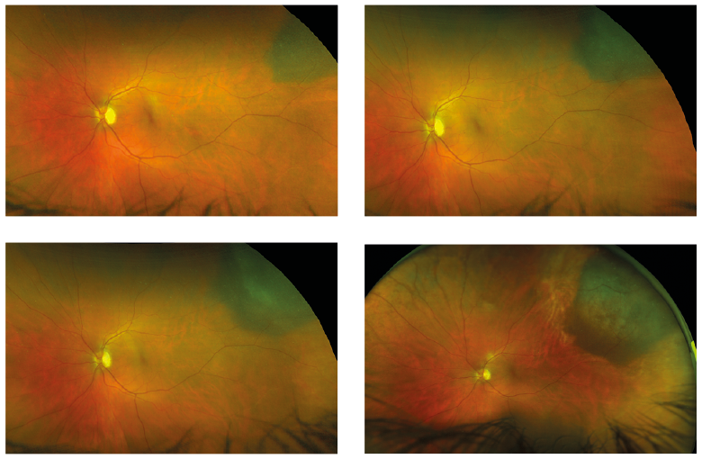
Choroidal Melanoma Recognizing Pathology Optos

Case 2 Bilateral Choroidal Melanoma Treated With Bilateral Plaque Download Scientific Diagram

Optometric Management Is It A Nevus Or Melanoma
Diagnosis Histopathologic And Genetic Classification Of Uveal Melanoma Intechopen
Choroidal Malignant Melanoma 58 Year Old Female With Pigmented Retinal Lesion And Exudative Retinal Detachment
Coms About Choroidal Melanoma
Nevus Or Melanoma How To Differentiate

The Small Fatal Choroidal Melanoma Study A Survey By The European Ophthalmic Oncology Group Sciencedirect

Full Text Optical Coherence Tomography Angiography In Choroidal Melanoma And Nev Opth

Choroidal Melanoma Uveal Melanoma Youtube
Spontaneous Regression Of Choroidal Melanoma Eye
Nevus Or Melanoma How To Differentiate
Choroidal Melanoma Europe American Academy Of Ophthalmology
Choroidal Melanoma Highland Retina Associates Terre Haute Indiana
Choroidal Nevus And Melanoma

Uveal Melanoma

Pdf Shaggy Photoreceptors With Small Choroidal Melanoma Semantic Scholar
Uveal Melanoma A Review Of The Literature Springerlink

Amelanotic Choroidal Melanoma Choroid Vessels Are Visible Through The Download Scientific Diagram
Choroidal Melanoma Practice Essentials Overview Pathophysiology
Q Tbn And9gcsg Dom Xwmf6rwuaxocyhfsinxieqv9mf79v73waxg Fqbwarm Usqp Cau
Choroidal Melanoma Retina Image Bank

Small Choroidal Melanoma A Ocular Fundus Photograph Shows Orange Download Scientific Diagram
Choroidal Melanoma Retina Orange County

Atypical Choroidal Melanoma Report Of 3 Cases

Choroidal Nevus Case Study

Amelanotic Choroidal Melanoma In 16 Month Old Child Canadian Journal Of Ophthalmology
Choroidal Melanoma Wills Eye Hospital

Small Juxtafoveal Melanoma Case 11 The Eye Cancer Network
What S The Risk Of Melanoma With A Choroidal Nevus Eyedolatry

Choroidal Melanoma Vs Retinoblastoma Ophthalmology Notes And Synopses Facebook
Uveal Melanoma Wikipedia
Gamma Knife Radiosurgery For Locally Recurrent Choroidal Melanoma Following Plaque Radiotherapy International Journal Of Retina And Vitreous Full Text

Choroidal Melanoma A Short Review With An Indian Perspective Pal Bp Garge S Khetan V Oman J Ophthalmol

Choroidal Melanoma October 19 Illinois Retina Associates

Choroidal Melanoma

Choroidal Melanoma The Eye Practice
Nonneoplastic Conditions That Can Simulate Posterior Uveal Melanoma And Other Intraocular Neoplasms Ento Key
Cureus Hidden Choroidal Melanoma Presented With Bullous Non Rhegmatogenous Retinal Detachment In A Myopic Asian Lady A Rare Manifestation
Imaging Advances For Choroidal Melanoma
Uveal Melanoma Abstract Europe Pmc
Choroidal Melanoma Retina Vitreous Surgeons Of Cny

Amelanotic Choroidal Melanoma The Lancet
.jpg)
Choroidal Melanoma Cancer Choroidal Melanoma Eye Cancer Treatment In Mumbai India

Choroidal Melanoma
Large Choroidal Malignant Melanoma Presenting As Neovascular Glaucoma

Pdf Choroidal Melanoma Natural History And Management Options Semantic Scholar
Choroidal Melanoma

Cancers Affecting The Retina Eye Disorders Merck Manuals Professional Edition
Choroidal Melanoma Is A Life Sentence

Choroidal Melanoma Case 3 Peripapillary Retina Image Bank
Choroidal Melanoma Europe American Academy Of Ophthalmology

Multimodal Imaging Of Suspicious Choroidal Neoplasms In A Primary Eye Care Clinic Kaur 17 Clinical And Experimental Optometry Wiley Online Library
Choroidal Melanoma New York Eye Cancer Center
Choroidal Melanoma Retina Vitreous Surgeons Of Cny
Optician
Q Tbn And9gctaklbm8rzf28jje8oyrbjigeysjbuorp07qnw 02lquo1rgibw Usqp Cau
Choroidal Melanoma Europe American Academy Of Ophthalmology

Choroid Melanoma Developed From Nevus Stoyukhina Ophthalmology Journal

Updated Ajcc Classification For Posterior Uveal Melanoma Retina Today

Choroidal Melanoma Case 4 Partly Amelanotic Retina Image Bank

Choroidal Melanoma Case 3 Color Af Fa Recognizing Pathology Optos

Endoresection Of Choroidal Melanoma British Journal Of Ophthalmology

Flat Choroidal Melanoma Masquerading As Central Serous Chorioretinopathy Higgins Tp Khoo Ct Magrath G Shields Cl Oman J Ophthalmol
Choroidal Melanoma Eye Life Vision Center


