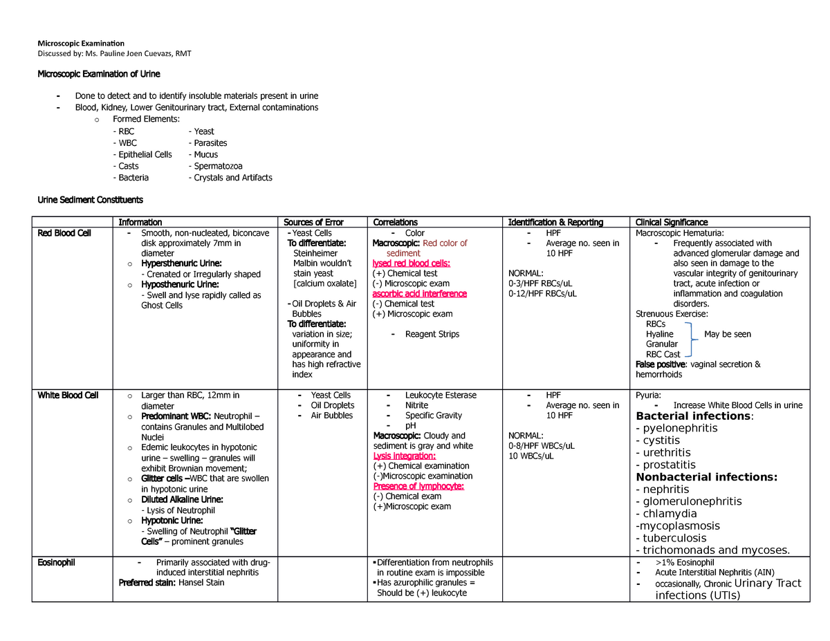
Yeast Cells In Urine Microscopy
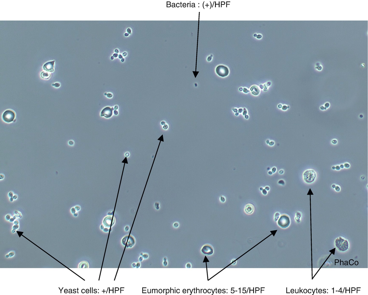
Artifacts PUS CELLS (WBC) The pus cells can enter in the urine anywhere from the glomerulus to the urethra Normal urine contain 23 pus cells per high power field (hpf) they are mostly Neutrophil and approximately 1012µ (micron) in diameter found in fresh voided urine Nuclear details may be lost with onset of cellular degeneration.
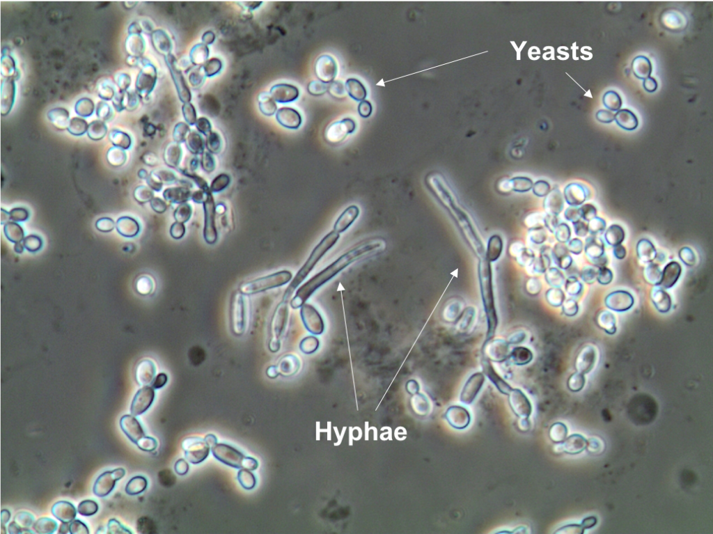
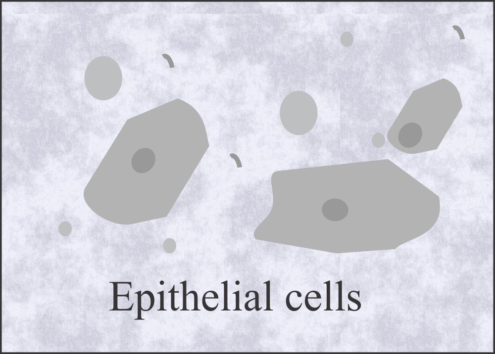
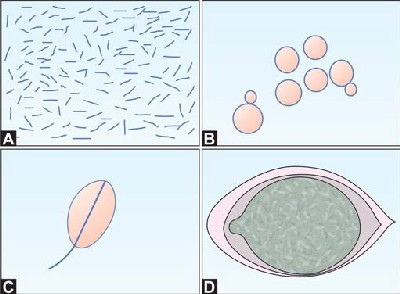
Yeast cells in urine microscopy. 44 Yeast cells Some studies found that the Iris iQ0 had a high false positive rate for yeast cell , , It was stated that yeast or crystals were not key elements for basic particle analysis, and could be eliminated by adjusting the corresponding thresholds in Iris iQ0 reports. Yeast Presence in Urine sediment by Light microscopy Active Part Description LP Yeast Fungi are eukaryotic microorganisms which manifest as yeasts, molds or a combination Yeasts start as individual cells that reproduce by budding Molds, which are long filaments or hyphae, grow by apical extension. Epithelial cells These cells normally slough in to the urine and can be seen in a urine sample They include transitional and squamous epithelial cells Casts Cylindrical structures produced in the kidney They are present in urine in certain disease states Bacteria Bacteria are common in urine However, the significant numbers may be indicative of an infection Yeast Yeast may either be contaminants or representative of true yeast infection.
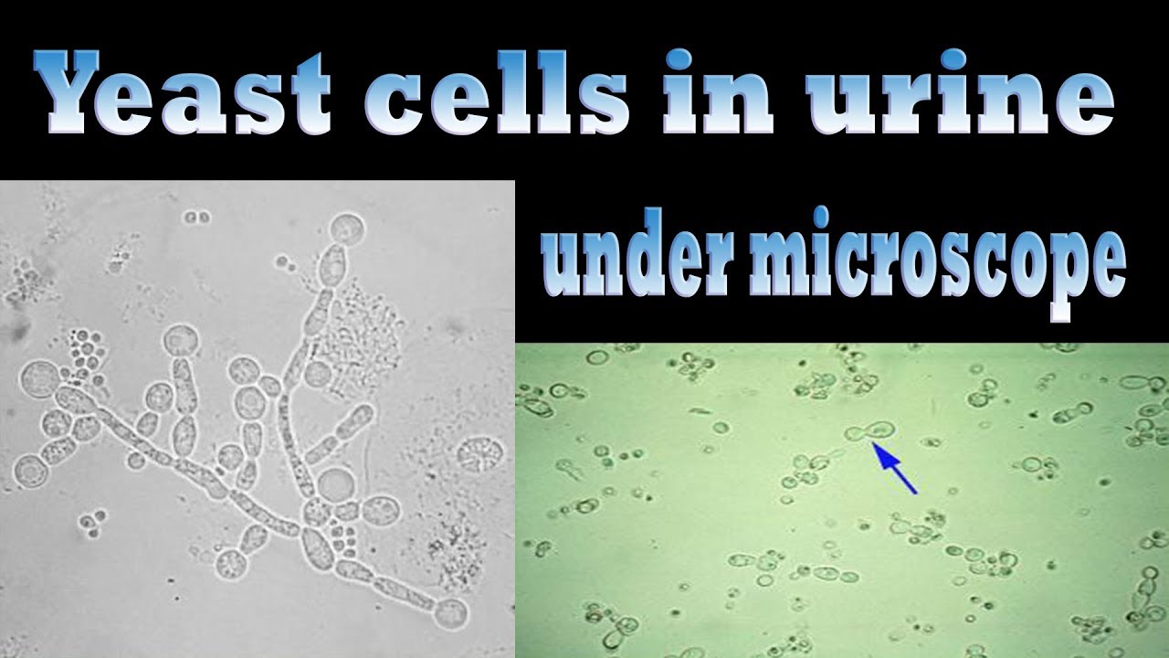
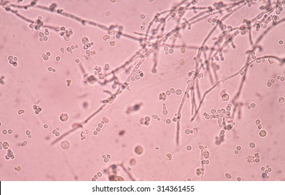
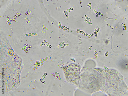
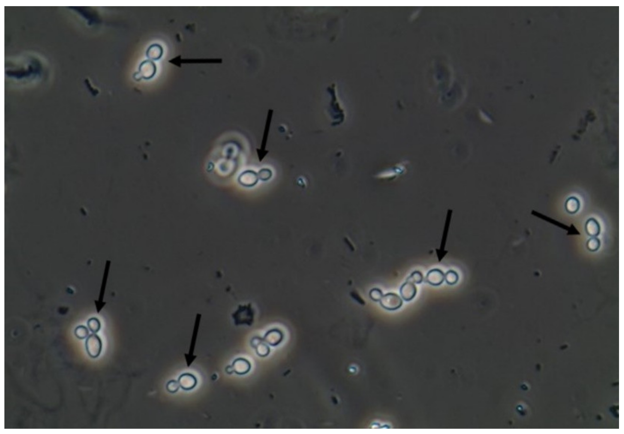
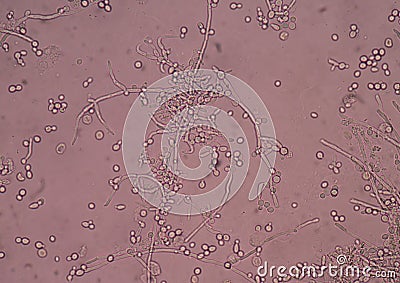
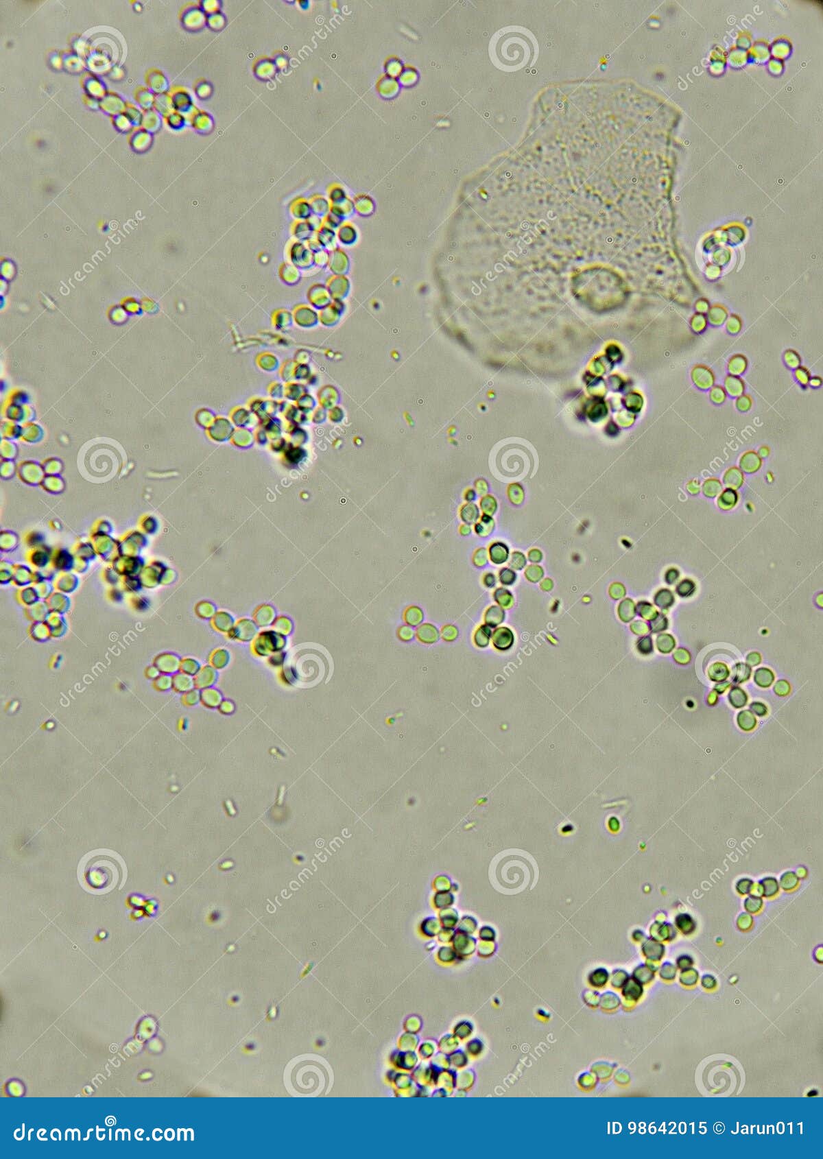
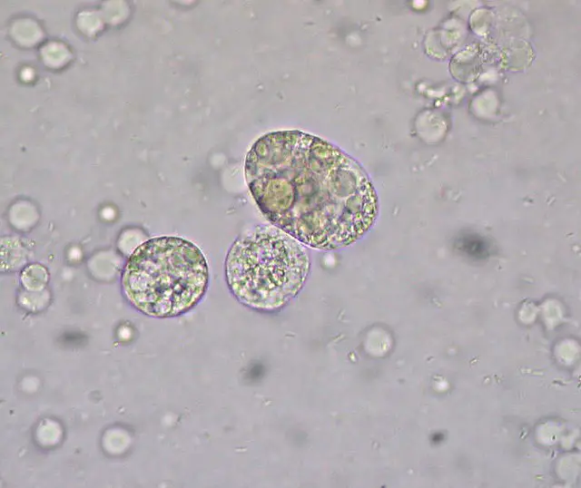
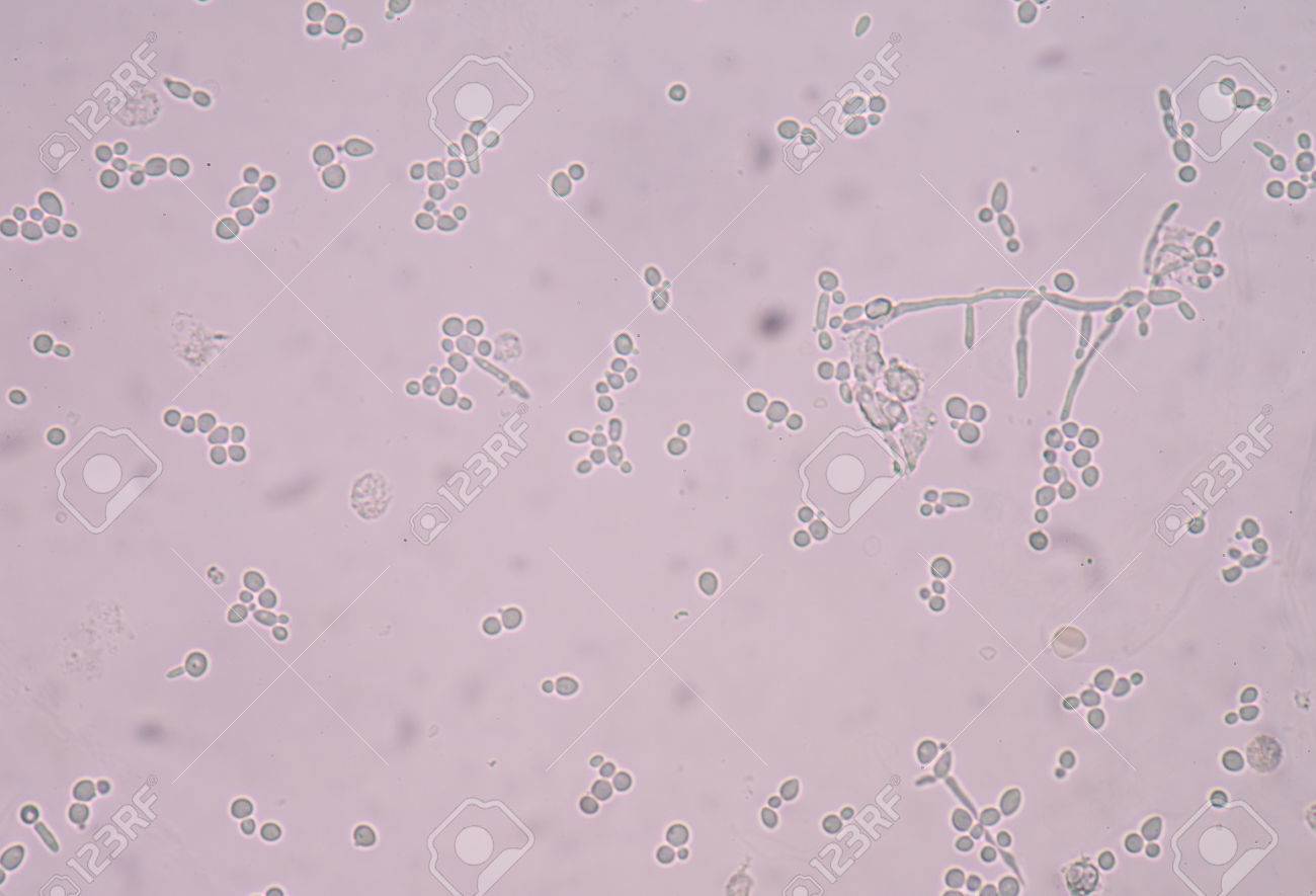
The Cell An Image Library™ is a public repository of images and videos These images have been reviewed and many have been annotated You will find that The Cell provides a large collection of yeast images and videos The Cell is funded by NIGMS Grand Opportunities grant RC2GM to the American Society for Cell Biology (ASCB). Yeast Yeast can appear as single cells or in the budding form, as indicated by the arrow in the top image The most common yeast found in urine is Candida albicans, which typically demonstrates budding forms The presence of budding forms helps to distinguish yeast from RBCs when urine sediment is viewed microscopically. In addition, red cell ghosts may simulate yeast The presence of dysmorphic RBC's in urine suggests a glomerular disease such as a glomerulonephritis Dysmorphic RBC's have odd shapes as a consequence of being distorted via passage through the abnormal glomerular structure.
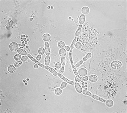
Start studying Microscopic Examination of Urine Learn vocabulary, terms, and more with flashcards, games, and other study tools most clinically significant urine epithelial cellsindicate tubular necrosis Yeast in urine characteristics nitrite negative, WBCs present. Yeast cells are more oval, show budding If identification is difficult, 2 preparation may be made and a few drops of acetic acid added to the one RBCs are lysed in the acidified preparation • 02 /HPF Normal ≥3/HPF in two of the three properly collected urine sample Microscopic hematuria. Photo about Branching budding yeast cells with pseudohyphae in urine fine with microscope Image of budding, microbiology, genetics.
Growing yeast cells for microscopy is easy, but you need to remember that you have to use actively growing culture;. Ensuring that a valid urine specimen is collected and carefully assessing patients for risk factors predisposing to disseminated candidiasis permit the stratification of cases into three Yeast in the urine solutions for a budding problem Clin Infect Dis 1995 Jan;(1)19 doi /clinids/11. Saccharomyces Cerevisiae Yeast Cells Under Microscope Written By MacPride Wednesday, September 6, 17 Add Comment Edit Yeast Cells Sem Photograph By Steve Gschmeissner Branching Budding Yeast Cells With Pseudohyphae In Urine Fine Gram Stain Saccharomyces Cerevisiae.
Yeast cells Bacteria and;. Some of the other staining techniques that can be used to differentiate certain urinary components include Sudan III, Sudan IV, and Oil Red O, which are used to stain fat a pink to red color;. Microscopic Examination of Urine Final Microscopic Examination Discussed by Ms Pauline Joen Cuevazs, RMT University Our Lady of Fatima University Course Medical Technology ((MLS)) Uploaded by Larae Zenal Academic year 18/19.
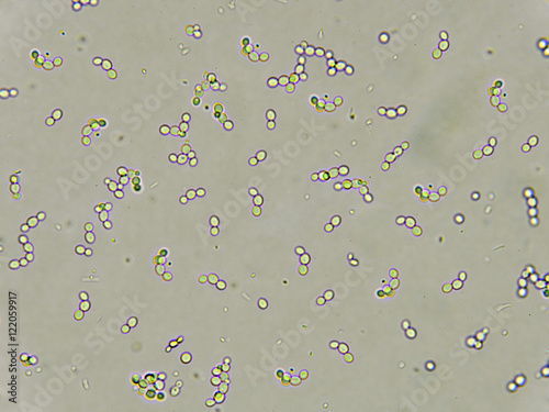
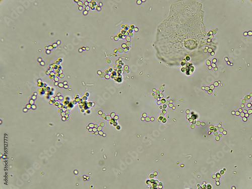
Yeast cells (Candida) These are round or oval structures of approx the same size as red blood cells In contrast to red cells, they show budding, are oval and more refractile, and are not soluble in 2% acetic acid Presence of Candida in urine may suggest immunocompromised state, vaginal candidiasis, or diabetes mellitus. And iodine, which can be used to stain starch granules and vegetable fibers a dark brown. Miscellaneous This category includes various cell forms and other structures that may be encountered in the urine sediment, such as yeast, bacteria, trichomonads, and fat globules When these are present, identify the cell or structure and report as few, moderate, or many per highpower field Report sperm as present in males only.
Yeast cells are more oval, show budding If identification is difficult, 2 preparation may be made and a few drops of acetic acid added to the one RBCs are lysed in the acidified preparation • 02 /HPF Normal ≥3/HPF in two of the three properly collected urine sample Microscopic hematuria. Ginal yeast infections affect most women at least once Common symptoms of a yeast infection include vaginal itching, burning, in this video you will s. Guide to interpretation of urine microscopy results There is no set normal range for cells in urine, as it is not possible to account for all patient types or conditions For example, a leukaemic or immunocompromised patient may have a low white cells count that may be regarded as normal in other patient groups etc.
Yeast and other fungal elements are common in stool Depending on the size and shape, they may be confused for a variety of helminth and protozoan species Figure C Nucleated red blood cell in a thin blood smear, stained with Giemsa There are several conditions which can cause a premature release of nucleated red blood cells into. They may be confused with yeast, pollen, starch, sperm heads, fat droplets and monohydrate calcium oxalate crystals Red cells can be differentiated because they are uniform, nonrefractile, lack a cell wall, have no internal structure and do not polarize. Ensuring that a valid urine specimen is collected and carefully assessing patients for risk factors predisposing to disseminated candidiasis permit the stratification of cases into three Yeast in the urine solutions for a budding problem Clin Infect Dis 1995 Jan;(1)19 doi /clinids/11.
Yeast and other fungal elements are common in stool Depending on the size and shape, they may be confused for a variety of helminth and protozoan species Figure C Nucleated red blood cell in a thin blood smear, stained with Giemsa There are several conditions which can cause a premature release of nucleated red blood cells into. Otherwise, many of your cells will be dead and useless Depending on your experiment, you can grow yeast in complete defined or complex media (YPD). Urine Under the Microscope As part of a urinalysis, the urine sediment is centrifuged and examined microscopically for crystals, casts, red blood cells, white bloods cells, and bacteria or yeast.
Describe the correct preparation of the urine sediment Recognize cells, casts, bacteria, yeast, crystals, and other structures that may be present in urine sediment Recognize artifacts in urine sediment and distinguish from significant findings Correlate microscopic findings with urine reagent strip findings. They may be confused with yeast, pollen, starch, sperm heads, fat droplets and monohydrate calcium oxalate crystals Red cells can be differentiated because they are uniform, nonrefractile, lack a cell wall, have no internal structure and do not polarize. Hey friends I'm medical laboratory scientistThis video has information about yeast cells in urine under microscope Microscopic photos(clear explanation) K.
Bacteria, Fungi, Candida, Yeast cells, or Parasites There are no bacteria, parasites or yeast cells in urine normally If these are present, it can mean you have an infection Presence of yeast in urine to be checked Squamous Cells The presence of squamous cells may mean that the sample is not as pure as it needs to be These cells do not mean there is a medical problem, but your doctor may ask that you give another urine sample. Yeast The most common yeast is candida in the urine This may be vaginal contamination due to vaginal infection These are yeast primarily candida seen in The patients with diabetes Because sugar is used as a source of energy for the growth of yeast These are also seen in immunocompromised patients Female with vaginal moniliasis. Eosin, which stains RBCs and helps distinguish them from yeast cells which will not pick up the stain;.
Describe the correct preparation of the urine sediment Recognize cells, casts, bacteria, yeast, crystals, and other structures that may be present in urine sediment Recognize artifacts in urine sediment and distinguish from significant findings Correlate microscopic findings with urine reagent strip findings. Yeast cells are normally not found in the urine of healthy individuals The term "yeast" is often used with infections caused by fungi Yeast cells in urine may be contaminants or represent a true yeast infection Interpret now "Complete Blood Count (CBC)" Interpret now "Comprehensive Metabolic Panel (CMP)" Contamination can be differentiated from colonization or infection by obtaining a new urine sample to verify funguria A carefully performed, cleanvoided, midstream sample is usually. Describe the correct preparation of the urine sediment Recognize cells, casts, bacteria, yeast, crystals, and other structures that may be present in urine sediment Recognize artifacts in urine sediment and distinguish from significant findings Correlate microscopic findings with urine reagent strip findings.
The finding of yeast cells in the urine is commonly associated with A Cystitis B Diabetes mellitus C Pyelonephritis D Liver disorders 30 The primary component of urinary mucus is A Bence Jones protein B Microalbumin C Tammhorsfall protein D Orthostatic protein 31 The majority of casts are formed in the A Proximal convoluted tubules. Microscopic Examination of Urine Final Microscopic Examination Discussed by Ms Pauline Joen Cuevazs, RMT University Our Lady of Fatima University Course Medical Technology ((MLS)) Uploaded by Larae Zenal Academic year 18/19. An increased number of WBCs seen in the urine under a microscope and/or positive test for leukocyte esterase may indicate an infection or inflammation somewhere in the urinary tract If also seen with bacteria (see below), they indicate a likely urinary tract infection.
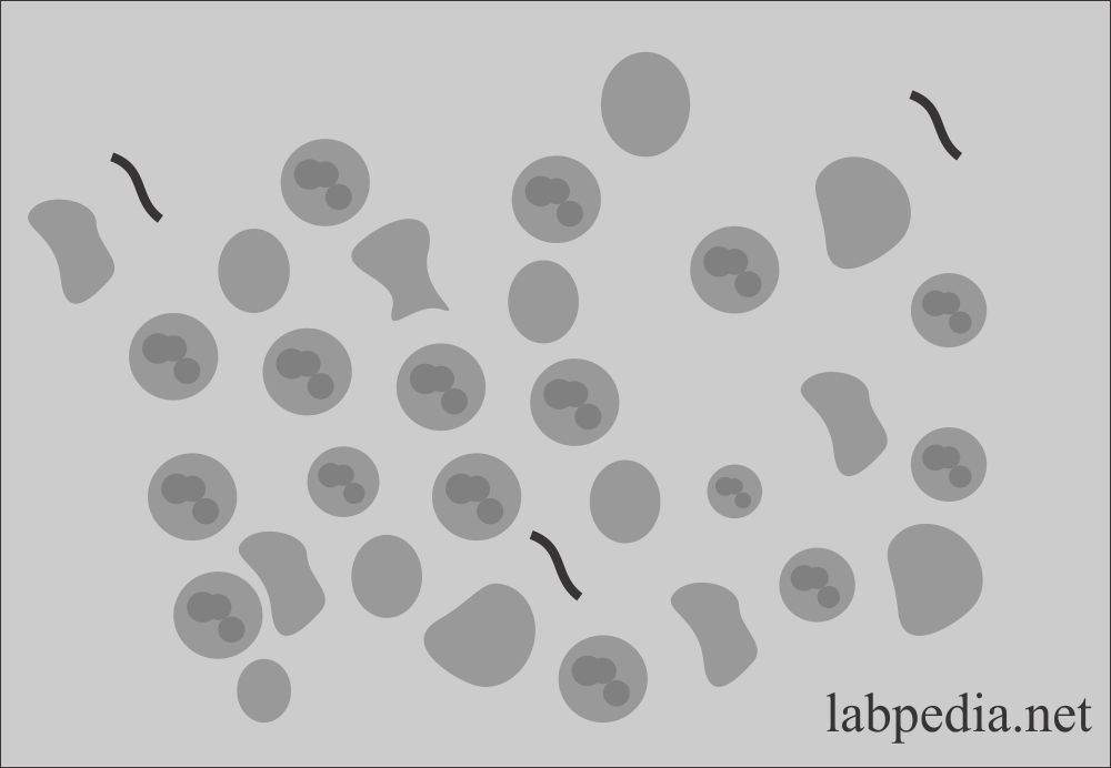
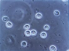
Guide to interpretation of urine microscopy results There is no set normal range for cells in urine, as it is not possible to account for all patient types or conditions For example, a leukaemic or immunocompromised patient may have a low white cells count that may be regarded as normal in other patient groups etc. Yeast Cells Yeast cells are smooth, colourless and usually ovoid cells These can vary in size and have double refractile walls They often show budding Unlike RBCs, they are insoluble in acid and alkaline They also will not stain with eosin These are mainly found in female urine due to skin and genital contamination. Yeast cells in urine oval structures that (usually are budding) reported as few, mod, many per high power field common seen yeast candidia alblicans (diabetic patients).
In addition, red cell ghosts may simulate yeast The presence of dysmorphic RBC's in urine suggests a glomerular disease such as a glomerulonephritis Dysmorphic RBC's have odd shapes as a consequence of being distorted via passage through the abnormal glomerular structure. A raised amount of epithelial cells in the urine are often the sign of a minor infection, such as a UTI or yeast infection Anyone with bothersome urinary symptoms should see a doctor for. The first thing yeast microscopy can be used for is a quick check with a simplest light microscope is whether your yeast culture is contaminated You don’t even need oil, just spot some cells on a glass slide At magnification x yeast cells are round, immobile and if they are from a growing culture budding.
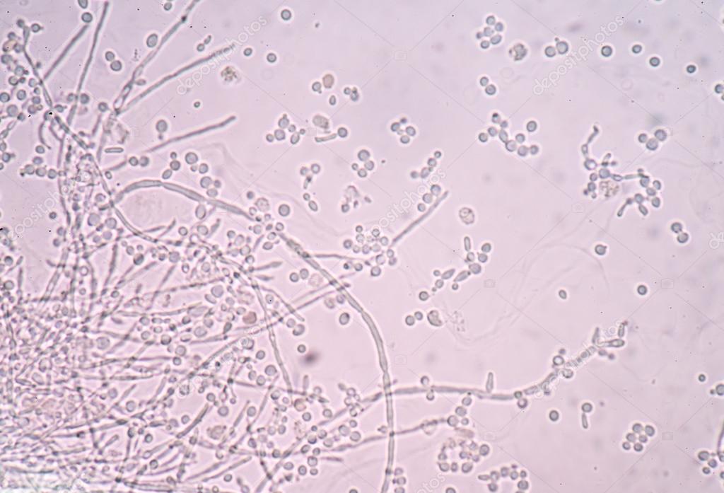
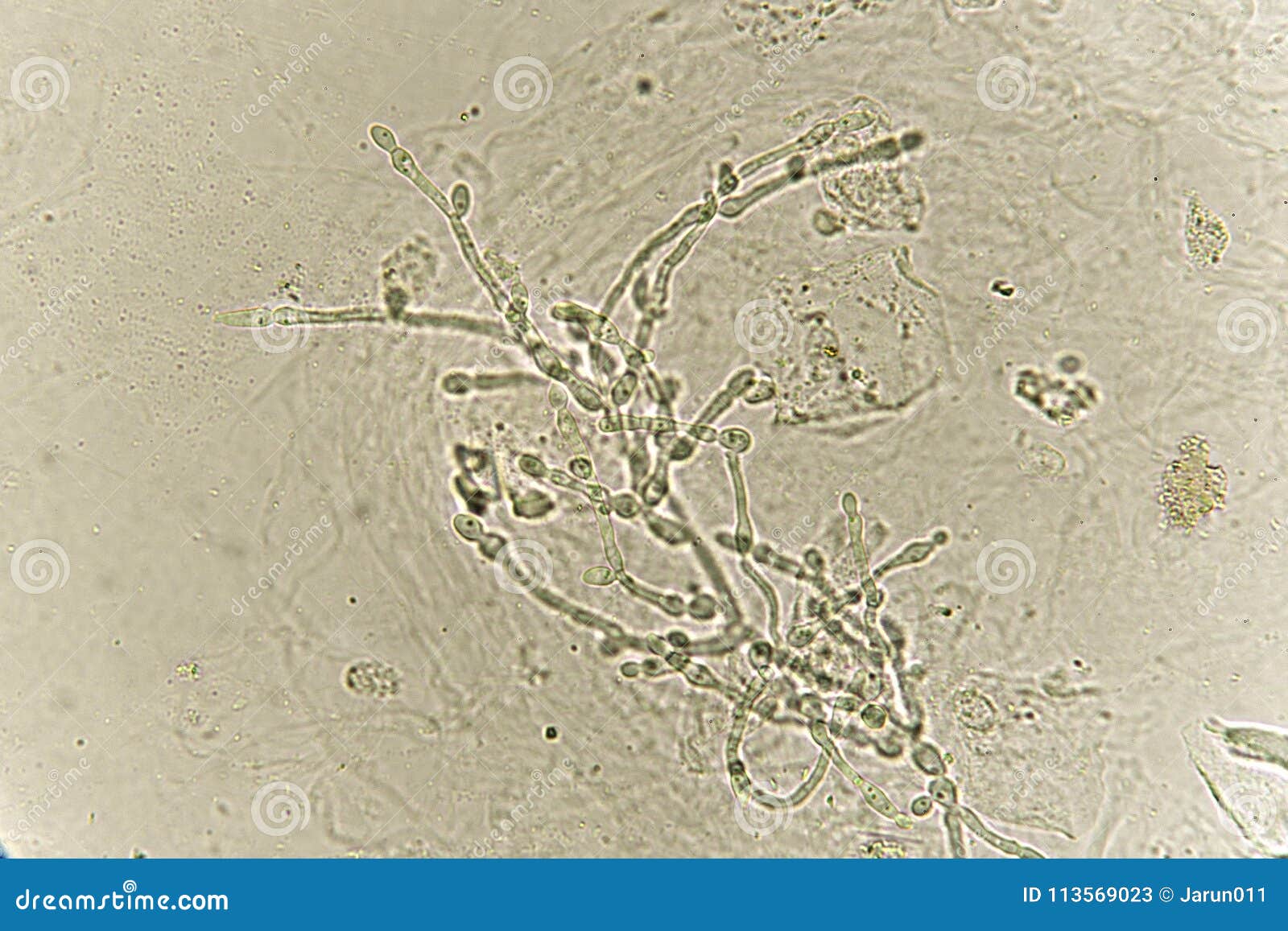
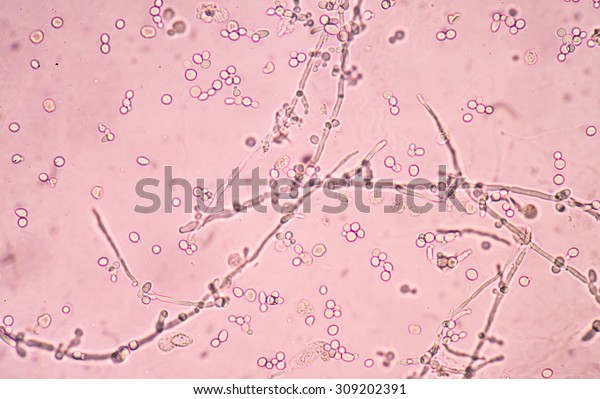
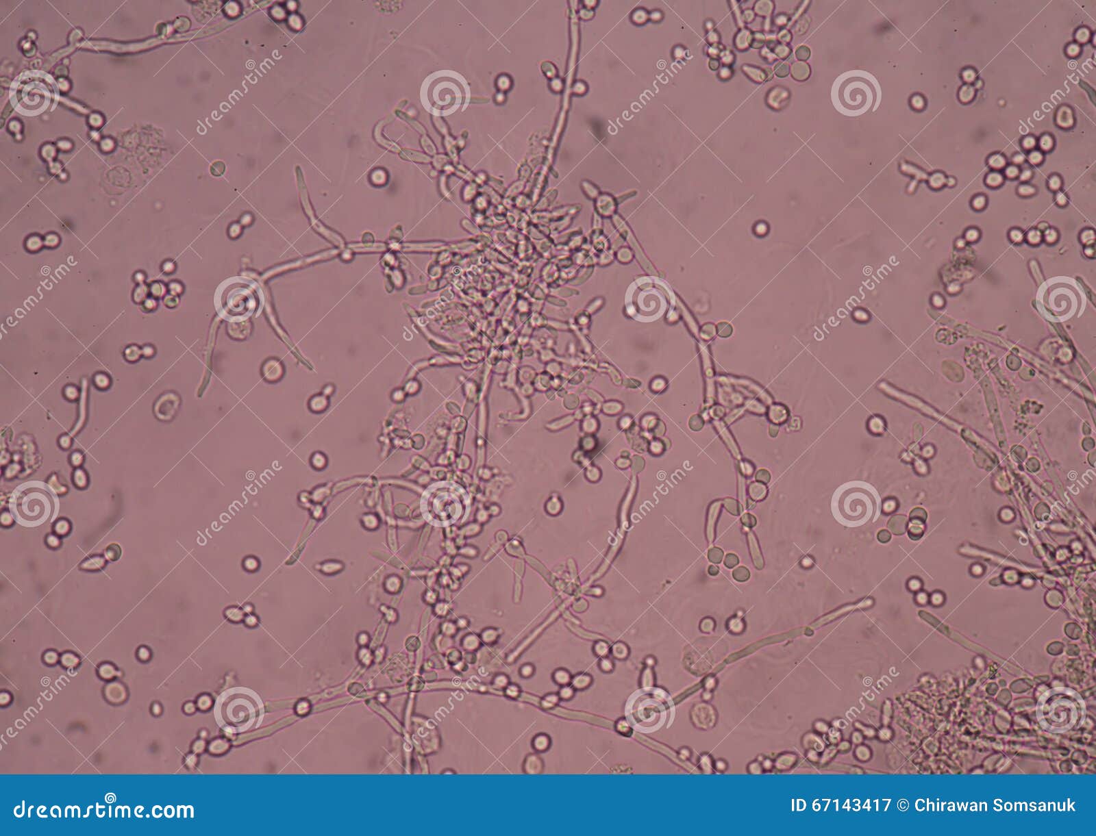
Yeasttohypha switching Although often referred to as dimorphic, C albicans is, in fact, polyphenic (often also referred to as pleomorphic) When cultured in standard yeast laboratory medium, C albicans grows as ovoid "yeast" cells However, mild environmental changes in temperature, CO 2, nutrients and pH can result in a morphological shift to filamentous growth. Dilute acetic acid will lyse red blood cells and aid in the distinction from yeast cells, oil droplets, or white blood cells Supravital stains may also be helpful Dysmorphic RBCs with spheroid surface protrusions are seen in renal/glomerular bleeding while eumorphic RBCs are seen in post renal bleeding Normally, red cells do not appear in the urine, although the presence of 12 RBC/HPF is usually not. Microscopic Evaluation • Start under low power (10x) objective • Scan the saline sample first for presence of •WBCs •Motile trichomonas •Yeast buds or pseudohyphae • Scan the KOH sample second for • Yeast buds or pseudohyphae • Other cells are lysed.
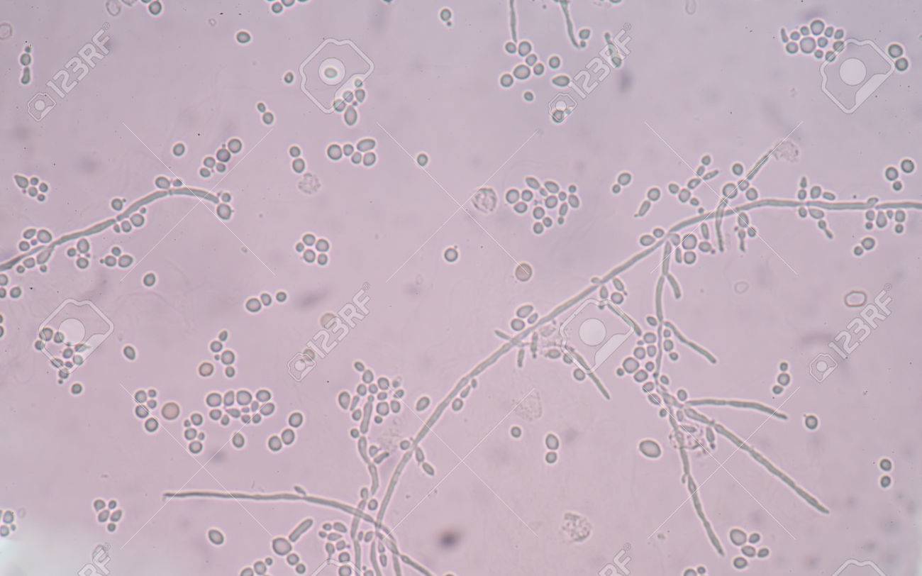
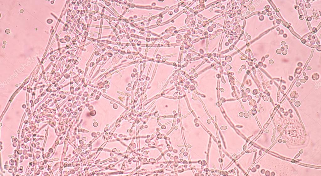
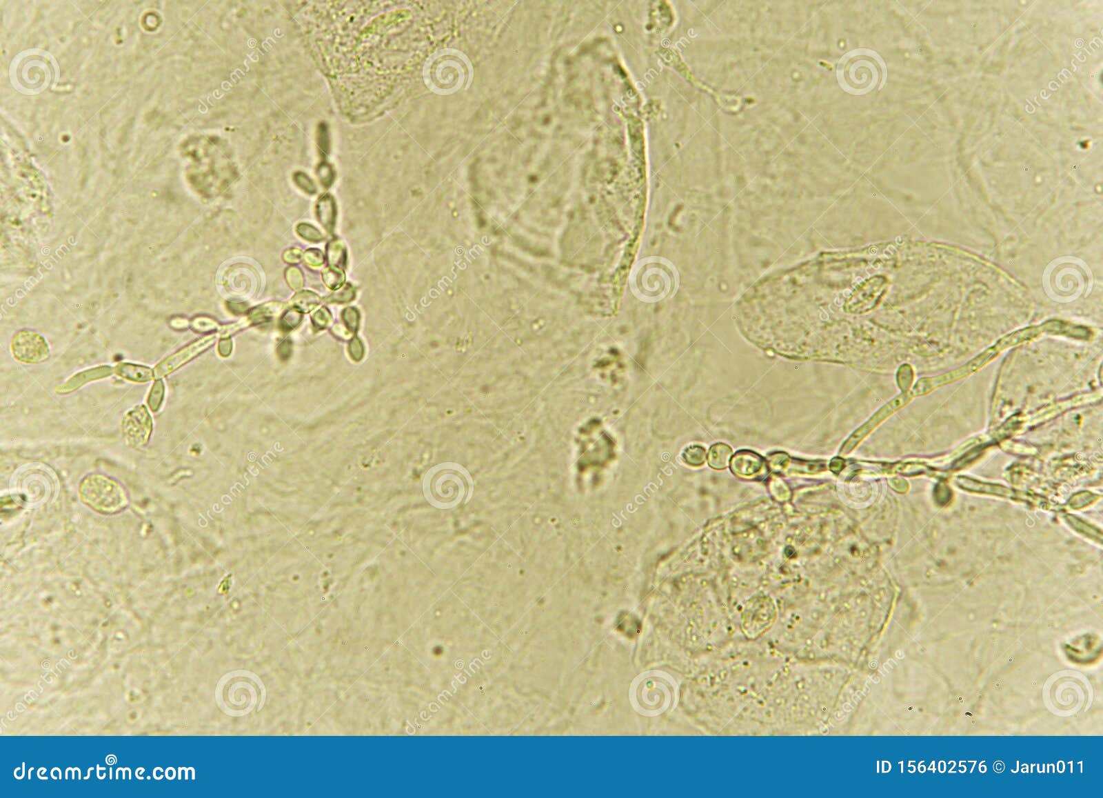
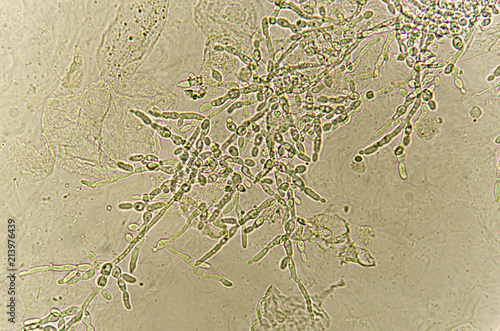
Photo about Pseudohyphae and budding yeast cells in patient urine, analyze by microscope Image of biology, analyzing, candidiasis. Bacteria, yeast cells, and parasites in urine sediment reflect genitourinary tract infection or contamination of external genitalia Yeast cells, which may be mistaken for red blood cells, are identified by their ovoid shape, lack of color, variable size, and frequently, signs of budding. Yeast Presence in Urine sediment by Light microscopy Active Part Description LP Yeast Fungi are eukaryotic microorganisms which manifest as yeasts, molds or a combination Yeasts start as individual cells that reproduce by budding Molds, which are long filaments or hyphae, grow by apical extension.
In urine, Candida albicans and other less commonly seen species, such as Candida parapsilosis and Candida tropicalis, will appear as budding yeasts, 4–10 μm in diameter, that often show formation of hyphal elements Smaller budding yeasts, only 2–4 μm in diameter, without any hyphal structures, are likely to be C glabrata. An increased number of WBCs seen in the urine under a microscope and/or positive test for leukocyte esterase may indicate an infection or inflammation somewhere in the urinary tract If also seen with bacteria (see below), they indicate a likely urinary tract infection. Cells Abnormal cells include renal tubular cells, caudate epithelial cells (from the renal pelvis), increased numbers of transitional epithelial cells, neoplastic cells, and inflammatory cells Organisms Bacteria, yeast, fungi, and parasite ova may be found in urine Biochemical Tests on Urine.
Presence of any of the following WBC’s, RBC’s, casts, crystals, yeast cells, bacteria etc is reported using sign depending on the number or amount found Their absence is reported as “nil” Urine is H 2 O containing the H 2 O – soluble waste products removed from the blood stream through the kidney Bacteriological examination of urine is done when the signs and symptoms of an infection or disease points to UTI’s, hypertension, and renal insufficiency. Yeast cells are normally not found in the urine of healthy individuals The term "yeast" is often used with infections caused by fungi Yeast cells in urine may be contaminants or represent a true yeast infection Interpret now "Complete Blood Count (CBC)". In a microscopic study, the urine sample is examined under a microscope to check for germs, cells, urine crystals or casts, etc Microorganisms (bacteria, fungi, yeast, trichomonads).
Yeast cells can be mistaken for RBCs Yeast cells are ovoid, rather than round, and they frequently contain buds which are smaller than themselves in size The doubly refractile border of the yeast cell tends to resemble the doughnut appearance of the red cell Yeast cells will not dissolve in 2% acetic acid, nor will they stain with eosin. In acidic pH *inorganic salts ppts in neutral & alkaline sol’n *except CaOx ppts in acidic & neutral urine Normal Crystal Seen in Acidic Urine Crystal Color & Shape Solubility Appearnce Uric Acid Yellowbrown Alkali (rhombic/4 Soluble sided/rosette) Amorphous Brick dust or Alkali and urates yellow brown heat granules Calcium Colorless. Yeast The most common yeast is candida in the urine This may be vaginal contamination due to vaginal infection These are yeast primarily candida seen in The patients with diabetes Because sugar is used as a source of energy for the growth of yeast These are also seen in immunocompromised patients Female with vaginal moniliasis.
# Pseudohyphae and budding yeast cells in patient urine Similar Images Add to Likebox # Petri dish with mixed of bacteria colonies Similar Images # budding yeast cell structure finding with microscope in laboratory Similar Images Add to Likebox # Fungus infection in white blood cells.
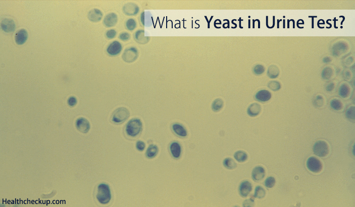
Yeast In Urine Test What Does Yeast Cells In Urine Mean
Q Tbn And9gctsb X Ridl2cnyseqdxdlwzvoaavammrcsbfhmddw Usqp Cau

Budding Yeast Cells Pseudohyphae Urine Sample Stock Photo Edit Now
Yeast Cells In Urine Microscopy のギャラリー
Urine Sediment Of The Month Microorganisms Renal Fellow Network

Photo Of Budding Yeast Cells With Id Royalty Free Image Stocklib
Http Webapps Cap Org Apps Docs Store Pub219 Urine Sample Pdf
Branching Budding Yeast Cells With Pseudohyphae In Urine Fine Wi Stock Photo C Toeytoey
Www Analyticon Diagnostics Com Downloads Flyer M2xfzr En Pdf
Haematology Microscopic Examination Of Urinary Sediment Miscellaneous Structures Free Medical Atlas

Madtechs Urine Microscopic Examination
Yeast Cells In Urine Under Microscope Microscopic Photos Clear Explanation Youtube

Photo Of Budding Yeast Cells In Urine Id Royalty Free Image Stocklib

Pseudohyphae Budding Yeast Cells Patient Urine Stock Photo Edit Now

Budding Yeast Cells Image Photo Free Trial Bigstock
Budding Yeast Cells With Pseudohyphae In Urine Sample Fine With Stock Photo Picture And Royalty Free Image Image
Fungal Urinary Tract Infection Encyclopedia
Www Analyticon Diagnostics Com Downloads Flyer M2xfzr En Pdf

Cme Slides Forum G B Fogazzi

Example Of Microscopic Urine Sediment Exam Results Medical Laboratories

Microscopic Urine Stock Photos And Images Agefotostock

Urinalysis
Education Kidney Org Sites Default Files Slides 11 Pdf

Vaginitis Diagnosis And Treatment American Family Physician
Epithelial Tissue With Bacteria Yeast Cells In Urine Sample Stock Photo Picture And Royalty Free Image Image

Yeast Cells Orange In Urine Sample Full Of Epithelial And Bacterial Cells X40 Medical Laboratory Technician Laboratory Technician Medical Laboratory

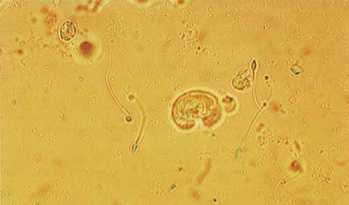
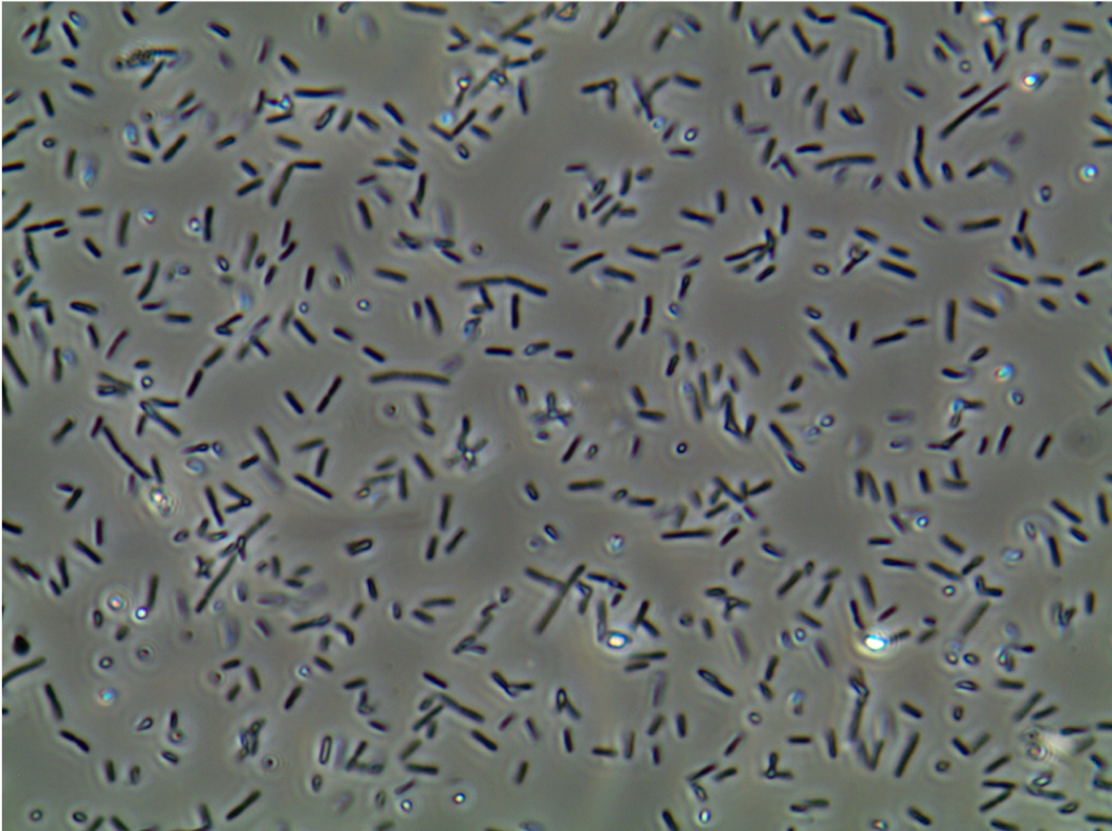
What Are These Picture Rod Shaped Organisms In Urine

Urinalysis Pictures Flashcards Quizlet

Budding Yeast Cells In Urine Stock Photo Download Image Now Istock

Micro 8 To Study Regulating Pathogens In Space Nasa

Budding Yeast Cells In Urine Stock Photo Picture And Royalty Free Image Image
1

Yeast Cells With Budding In Urine Microscopy At 40x Youtube
Branching Budding Yeast Cells With Pseudohyphae In Urine Fine W Stock Photo C Toeytoey
Yeast Microscope Hd Stock Images Shutterstock

Budding Yeast Cells With Pseudohyphae In Urine Sample Fine With Stock Photo Picture And Royalty Free Image Image

Microscopic Examination Of Urine Showing Pus Cells Budding Yeast Cells Download Scientific Diagram

Urinary Tract Infection Wikipedia
Microscopic Urine Sediment Analysis And Findings Springerlink

Cells Found In Urine Sediment Aladdin Creations
Budding Yeast Cells In Patient Urine Urinary Tract Infections Analyze By Microscope Contrast Adjustment Buy This Stock Photo And Explore Similar Images At Adobe Stock Adobe Stock

Red And White Blood Cells And Yeast In Urine Sediment Medical Laboratories

Budding Yeast Cells In Urine Stock Photo Picture And Royalty Free Image Image

Budding Yeast Cells Budding Yeast In Urine Budding Yeast Under Microscope Youtube

Urine Test

Pseudohyphae And Budding Yeast Cells In Urine Stock Photo Download Image Now Istock
Q Tbn And9gcsnjtepdlumoyg2gtcsa4xev3 Q2tiqndbkkddmalriaqvrjxmj Usqp Cau
Fungal Urinary Tract Infection Encyclopedia
Urine Analysis Part 6 Urine Microscopic Examination And Interpretations Labpedia Net
Microscopic Urine Sediment Analysis And Findings Springerlink
Www Analyticon Diagnostics Com Downloads Flyer M2xfzr En Pdf
Budding Yeast Cells In Urine Analyze By Microscope Buy This Stock Photo And Explore Similar Images At Adobe Stock Adobe Stock

Cme Slides Forum G B Fogazzi
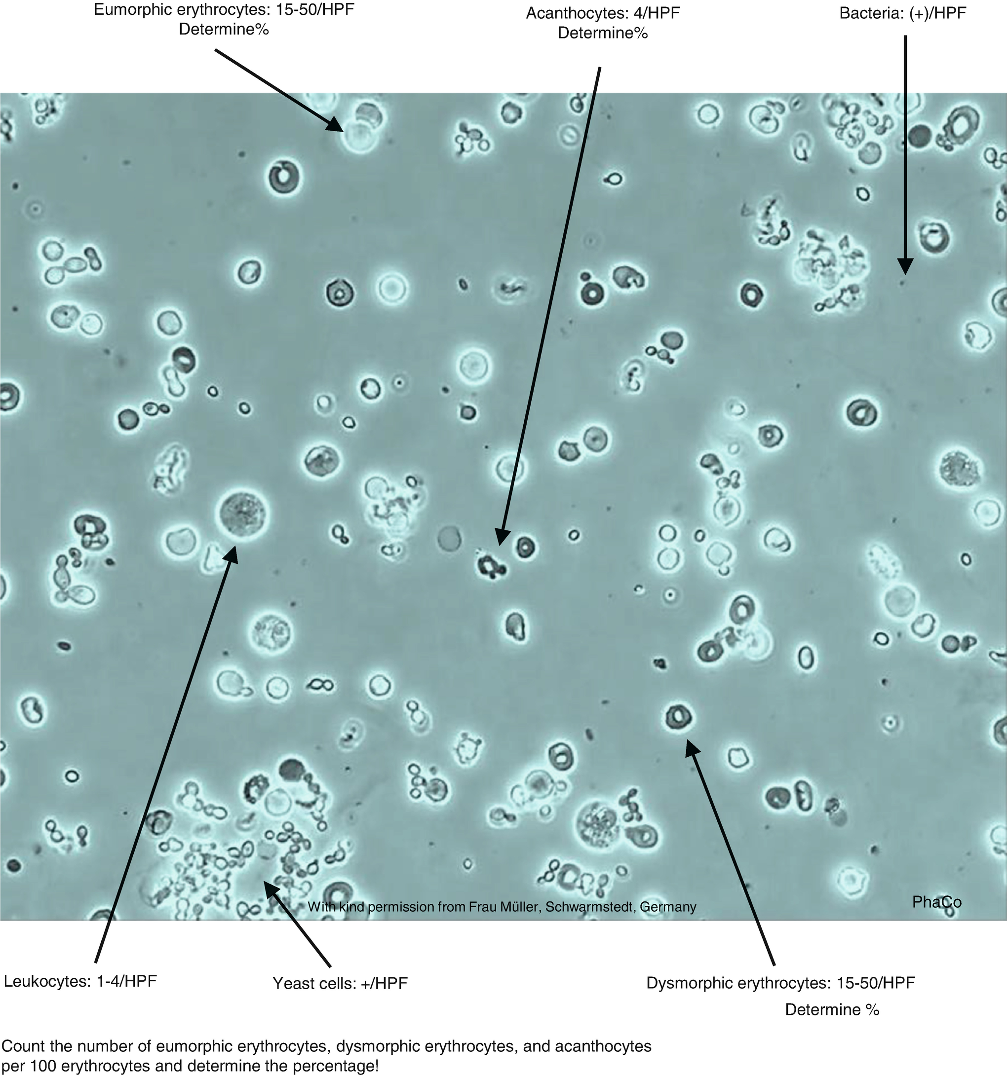
Urine Analysis Part 6 Urine Microscopic Examination And Interpretations Labpedia Net

Yeast Cells In Urine Urinal Yeast Cell

Long Chain Bacteria Yeast Cell Budding Pseudohyphae Pus Cells In Urine Youtube

Urinalysis

Epithelial Tissue With Bacteria Cells Yeast Cells Epithelial Tissue With Bacteria Yeast Cells In Urine Sample Under Canstock

Budding Yeast Cells In Urine Stock Photo Download Image Now Istock
Procedures For Complete Urinalysis Confirmation Testing Springerlink
Fungal Urinary Tract Infection Encyclopedia
Jof Free Full Text Urine Sediment Findings And The Immune Response To Pathologies In Fungal Urinary Tract Infections Caused By Candida Spp

Haematology Microscopic Examination Of Urinary Sediment Cells Free Medical Atlas
Http Www Api Pt Com Reference Commentary 14bscope2 Pdf
Pseudohyphae And Budding Yeast Cells In Patient Urine Stock Image Image Of Fungi Antibiotic

Pseudohyphae And Budding Yeast Cells In Patient Urine Stock Image Image Of Infection Bacteria
Http Www Api Pt Com Reference Commentary 14bscope2 Pdf
Budding Yeast Cells Pseudohyphae Urine Sample Stock Photo Edit Now
Http Webapps Cap Org Apps Docs Store Pub219 Urine Sample Pdf
Urine Analysis Part 6 Urine Microscopic Examination And Interpretations Labpedia Net
Urine Sediment Of The Month Dysmorphic Red Blood Cells Blebs Spikes Renal Fellow Network

Budding Yeast Cells Bacteria Patient Urine Stock Photo Edit Now
Urine Sediment Of The Month Microorganisms Renal Fellow Network

Yeast In Urine Sediment Medical Laboratories

Branching Budding Yeast Cells With Pseudohyphae In Urine Sample Stock Photo Picture And Royalty Free Image Image
Q Tbn And9gcq74xpy Ew Q4p6elrggql4kaeqlxlopd8nyqncp1bmlzckzuyo Usqp Cau
Pseudohyphae And Budding Yeast Cells In Patient Urine Stock Photo Image Of Laboratory Bacteria
Pseudohyphae And Budding Yeast Cells In Patient Urine Urinary Tract Infections Analyze By Microscope Buy This Stock Photo And Explore Similar Images At Adobe Stock Adobe Stock

Branching Budding Image Photo Free Trial Bigstock

Pin On Lab Expert

Budding Yeast Cells Stock Photo Download Image Now Istock

Budding Yeast Cells Stock Photo Download Image Now Istock

Microscopic Analysis Of Urine Faculty Of Medicine Masaryk University
Www Wjpmr Com Download Article Pdf
Budding Yeast Cells With Pseudohyphae In Urine Sample Royalty Free Stock Photo Cartoondealer Com
Budding Yeast Cells In Urine Analyze By Microscope Buy This Stock Photo And Explore Similar Images At Adobe Stock Adobe Stock
Examination Of The Urinary Sediment Veterian Key
Budding Yeast Cells In Patient Urine Urinary Tract Infections Analyze By Microscope Contrast Adjustment Buy This Stock Photo And Explore Similar Images At Adobe Stock Adobe Stock
Budding Yeast Cells Stock Image Image Of Epithelium

Budding Yeast Cells In Urine Stock Photo Download Image Now Istock

Pseudohyphae And Budding Yeast Cells In Urine Stock Image Image Of Candida Analyzing

Urinalysis Sediment Cells Casts Crystals Flashcards Quizlet
Budding Yeast Cells With Pseudohyphae In Urine Sample Stock Photo Megapixl
Microscopic Examination Of Urine
Microscopy Culture And Sensitivity Urine Sputum Blood And Stool

Direct Microscopy Of The Urinary Sediment Showing Yeast Cells 4009 Download Scientific Diagram
Budding Yeast Cells With Pseudohyphae In Urine Sample Fine With Stock Photo Picture And Royalty Free Image Image

Pseudohyphae And Budding Yeast Cells In Urine Stock Image Image Of Candida Analyzing
Academic Oup Com Ajcp Article Pdf 112 1 25 Ajcpath112 0025 Pdf
Microscopic Examination Of Urine Final Studocu
Yeast Infection Candidiasis My Doctor Online

Budding Yeast Cell In Urine Microscopy Pathology Lab Sajid Facebook
Www Analyticon Diagnostics Com Downloads Flyer M2xfzr En Pdf
Urinary Tract Infection Wikipedia

Urine Microscopic Ideas Medical Laboratory Medical Laboratory Science Medical Laboratory Scientist


